Role of Diffusion and Perfusion Imaging Measurements in Grading of Glioma and Quantitative Assessment of Temporal Change in These Measurements in Glioma Patients Undergoing Radiotherapy
Article Sidebar
Main Article Content
Abstract
Introduction: Conventional neuro imaging of brain tumors is anatomy based and differentiates low grade (LGG) and high grade glioma (HGG) based on pattern recognition which at times may be difficult to comprehend. Functional MRI incorporates morphologic abnormality with physiologic alterations. Quantification of ADC (apparent diffusion coefficient) and rCBV (relative cerebral blood volume) values using functional MRI have been explored to characterize these tumors. The present study purposes to determine the temporal change in diffusion (ADC) and perfusion (rCBV) in glioma patients treated by radiotherapy.
Material and Methods: 3Tesla MRI diffusion & perfusion imaging was done postoperatively before radiotherapy (RT), during RT at 4th week & subsequently on follow-up on 18 glioma patients (LGG=5 and HGG=13). Quantification of ADCminimum (ADCmin) and rCBVmaximum (rCVBmax) value was done and the temporal changes of these parameters with RT were evaluated.
Results: In pre-RT MRI, the mean ADCmin for LGG was significantly higher in comparison to HGG (mean ± SD, 1.10 ± 0.11; 0.83 ± 0.13, respectively, all values x 10-3 mm2 sec-1; t-test p=0.00). The mean ADCmin for LGG during RT increased compared to baseline value and thereafter remained stable at the first follow-up imaging, although the difference was non-significant (p=.33 and 0.8, respectively), while for HGG, mean ADCmin significantly increased during RT from baseline value and consistently increased at the time of follow-up imaging (p=.00, p=.01, respectively).
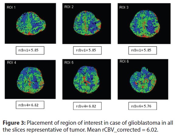
The rCVBmax for LGG (1.98 ± 0.70) was significantly lower in comparison to HGG (5.07 ± 0.04). In both LGG and HGG the rCVBmax decreased during RT from baseline pre-RT values and remained constant at the follow-up. The mean difference in rCVBmax values was significant (LGG: p = 0.00 & 0.03; HGG: p = 0.00 & 0.002, respectively).
Conclusion: There is a change in diffusion (ADCmin) and perfusion (rCVBmax) parameters in glioma patients treated by radiotherapy with values differing based on the grade of tumor, and these measurements shows a temporal change in their values with radiation response.
Article Details

This work is licensed under a Creative Commons Attribution-NonCommercial-ShareAlike 4.0 International License.
The Journals licensing terms of CC-BY-NC-SA 4.0 speaks that you are free to Share (copy and redistribute the material in any medium or format), Adapt (remix, transform, and build upon the material) under proper terms of Attribution (You must give appropriate credit, provide a link to the license, and indicate if changes were made. You may do so in any reasonable manner, but not in any way that suggests the licensor endorses you or your use.), NonCommercial (You may not use the material for commercial purposes.), ShareAlike (If you remix, transform, or build upon the material, you must distribute your contributions under the same license as the original.) and No additional restrictions (You may not apply legal terms or technological measures that legally restrict others from doing anything the license permits.)
References
.