Neurological Implications of COVID-19: From Cognitive Decline to Blood Biomarkers COVID-19 and brain
Article Sidebar
Main Article Content
Abstract
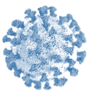
The COVID-19 pandemic has endured since 2019. The severe acute respiratory syndrome coronavirus 2 (SARS-CoV-2) affects a variety of organs, including the brain. Neurological complications after post-COVID-19 sequelae have been recognized in several reports. There is a plethora of evidence that COVID-19 sufferers have neurological, cognitive, and emotional problems. Among those infected with SARS-CoV-2, acute neurological symptoms like cognitive decline, neuroinflammation, cognitive decline, brain stroke, and loss of smell, are frequent direct outcomes. Three months after being infected with SARS-CoV-2, neurological abnormalities were recorded in up to 55% of COVID-19 patients in the hospital. The SARS-COV-2 virus' mutability and propensity for direct central nervous system (CNS) damage underscore the pressing need for technology to identify, manage, and treat brain injury in COVID-19 patients. This review explores the direct and indirect impacts of COVID-19 on the CNS and delves into the underlying causes and risk factors contributing to the deterioration of individuals' mental well-being and the COVID-19 pandemic. Furthermore, we also discussed the diagnostic blood biomarkers (BBs) for brain injury in patients with COVID-19.
Article Details

This work is licensed under a Creative Commons Attribution-NonCommercial-ShareAlike 4.0 International License.
The Journals licensing terms of CC-BY-NC-SA 4.0 speaks that you are free to Share (copy and redistribute the material in any medium or format), Adapt (remix, transform, and build upon the material) under proper terms of Attribution (You must give appropriate credit, provide a link to the license, and indicate if changes were made. You may do so in any reasonable manner, but not in any way that suggests the licensor endorses you or your use.), NonCommercial (You may not use the material for commercial purposes.), ShareAlike (If you remix, transform, or build upon the material, you must distribute your contributions under the same license as the original.) and No additional restrictions (You may not apply legal terms or technological measures that legally restrict others from doing anything the license permits.)
Funding data
-
Department of Biotechnology, Ministry of Science and Technology, India
Grant numbers BT/IN/Indo-US/Foldscope/39/2015 -
Indian Council of Medical Research
Grant numbers 3/1/2/190/Neuro/2021-NCD-I
References
WHO. Coronavirus disease (COVID-2019) situation reports. 2020. https://www.who.int/emergencies/diseases/novel-coronavirus-2019/situationreports. Accessed 22 Mar 2020
Zhu N, Zhang D, Wang W, Li X, Yang B, Song J, Zhao X, Huang B, Shi W, Lu R, Niu P. A novel coronavirus from patients with pneumonia in China, 2019. New England journal of medicine. 2020 Feb 20;382(8):727-33.
Lu R, Zhao X, Li J, Niu P, Yang B, Wu H, Wang W, Song H, Huang B, Zhu N, Bi Y. Genomic characterisation and epidemiology of 2019 novel coronavirus: implications for virus origins and receptor binding. The lancet. 2020 Feb 22;395(10224):565-74.
Su S, Wong G, Shi W, Liu J, Lai AC, Zhou J, Liu W, Bi Y, Gao GF. Epidemiology, genetic recombination, and pathogenesis of coronaviruses. Trends in microbiology. 2016 Jun 1;24(6):490-502.
Du L, He Y, Zhou Y, Liu S, Zheng BJ, Jiang S. The spike protein of SARS-CoV—a target for vaccine and therapeutic development. Nature Reviews Microbiology. 2009 Mar;7(3):226-36.
Gallagher TM, Buchmeier MJ. Coronavirus spike proteins in viral entry and pathogenesis. Virology. 2001 Jan 20;279(2):371-4.
Simmons G, Zmora P, Gierer S, Heurich A, Pöhlmann S. Proteolytic activation of the SARS-coronavirus spike protein: cutting enzymes at the cutting edge of antiviral research. Antiviral research. 2013 Dec 1;100(3):605-14.
Song W, Gui M, Wang X, Xiang Y. Cryo-EM structure of the SARS coronavirus spike glycoprotein in complex with its host cell receptor ACE2. PLoS pathogens. 2018 Aug 13;14(8):e1007236.
Li F, Li W, Farzan M, Harrison SC. Structure of SARS coronavirus spike receptor-binding domain complexed with receptor. Science. 2005 Sep 16;309(5742):1864-8.
Belouzard S, Chu VC, Whittaker GR. Activation of the SARS coronavirus spike protein via sequential proteolytic cleavage at two distinct sites. Proceedings of the National Academy of Sciences. 2009 Apr 7;106(14):5871-6.
Millet JK, Whittaker GR. Host cell proteases: critical determinants of coronavirus tropism and pathogenesis. Virus research. 2015 Apr 16;202:120-34.
Simmons G, Gosalia DN, Rennekamp AJ, Reeves JD, Diamond SL, Bates P. Inhibitors of cathepsin L prevent severe acute respiratory syndrome coronavirus entry. Proceedings of the National Academy of Sciences. 2005 Aug 16;102(33):11876-81.
Kishimoto TK, Larson RS, Corbi AL, Dustin ML, Staunton DE, Springer TA. The leukocyte integrins. Advances in immunology. 1989 Jan 1;46:149-82.
Larson RS, Springer TA. Structure and function of leukocyte integrins. Immunological reviews. 1990 Apr;114(1):181-217.
Bunting M. Harris ES, McIntyre TM, Prescott SM, and Zimmerman GA. Leukocyte adhesion deficiency syndromes: adhesion and tethering defects involving beta.;2:30-5.
Diamond MS, Springer TA. The dynamic regulation of integrin adhesiveness. Current Biology. 1994 Jun 1;4(6):506-17.
Imhof BA, Dunon D. Leukocyte migration and adhesion. Advances in immunology. 1995 Jan 1;58:345-416.
Bauer Jr TR, Hickstein DD. Gene therapy for leukocyte adhesion deficiency. Current opinion in molecular therapeutics. 2000 Aug 1;2(4):383-8.
Zhou P, Yang XL, Wang XG, Hu B, Zhang L, Zhang W, Si HR, Zhu Y, Li B, Huang CL, Chen HD. A pneumonia outbreak associated with a new coronavirus of probable bat origin. nature. 2020 Mar;579(7798):270-3.
Meinhardt J, Radke J, Dittmayer C, Franz J, Thomas C, Mothes R, Laue M, Schneider J, Brünink S, Greuel S, Lehmann M. Olfactory transmucosal SARS-CoV-2 invasion as a port of central nervous system entry in individuals with COVID-19. Nature neuroscience. 2021 Feb;24(2):168-75.
Daniels BP, Holman DW, Cruz-Orengo L, Jujjavarapu H, Durrant DM, Klein RS. Viral pathogen-associated molecular patterns regulate blood-brain barrier integrity via competing innate cytokine signals. MBio. 2014 Oct 31;5(5):10-128.
Al-Dalahmah O, Thakur KT, Nordvig AS, Prust ML, Roth W, Lignelli A, Uhlemann AC, Miller EH, Kunnath-Velayudhan S, Del Portillo A, Liu Y. Neuronophagia and microglial nodules in a SARS-CoV-2 patient with cerebellar hemorrhage. Acta Neuropathologica Communications. 2020 Dec;8(1):1-7.
Liddelow SA, Guttenplan KA, Clarke LE, Bennett FC, Bohlen CJ, Schirmer L, Bennett ML, Münch AE, Chung WS, Peterson TC, Wilton DK. Neurotoxic reactive astrocytes are induced by activated microglia. Nature. 2017 Jan 26;541(7638):481-7.
Vasek MJ, Garber C, Dorsey D, Durrant DM, Bollman B, Soung A, Yu J, Perez-Torres C, Frouin A, Wilton DK, Funk K. A complement–microglial axis drives synapse loss during virus-induced memory impairment. Nature. 2016 Jun 23;534(7608):538-43.
Beyrouti R, Adams ME, Benjamin L, Cohen H, Farmer SF, Goh YY, Humphries F, Jäger HR, Losseff NA, Perry RJ, Shah S. Characteristics of ischaemic stroke associated with COVID-19. Journal of Neurology, Neurosurgery & Psychiatry. 2020 Aug 1;91(8):889-91.
Bowles L, Platton S, Yartey N, Dave M, Lee K, Hart DP, MacDonald V, Green L, Sivapalaratnam S, Pasi KJ, MacCallum P. Lupus anticoagulant and abnormal coagulation tests in patients with Covid-19. New England Journal of Medicine. 2020 Jul 16;383(3):288-90.
Wang Z, Yang Y, Liang X, Gao B, Liu M, Li W, Chen Z, Wang Z. COVID-19 associated ischemic stroke and hemorrhagic stroke: incidence, potential pathological mechanism, and management. Frontiers in Neurology. 2020 Oct 27;11:571996.
Becker RC. COVID-19 update: Covid-19-associated coagulopathy. Journal of thrombosis and thrombolysis. 2020 Jul;50(1):54-67.
Mao L, Jin H, Wang M, Hu Y, Chen S, He Q, Chang J, Hong C, Zhou Y, Wang D, Miao X. Neurologic manifestations of hospitalized patients with coronavirus disease 2019 in Wuhan, China. JAMA neurology. 2020 Jun 1;77(6):683-90.
Chen L, Li Q, Zheng D, Jiang H, Wei Y, Zou L, Feng L, Xiong G, Sun G, Wang H, Zhao Y. Clinical characteristics of pregnant women with Covid-19 in Wuhan, China. New England Journal of Medicine. 2020 Jun 18;382(25):e100.
Abdullahi A, Candan SA, Abba MA, Bello AH, Alshehri MA, Afamefuna Victor E, Umar NA, Kundakci B. Neurological and musculoskeletal features of COVID-19: a systematic review and meta-analysis. Frontiers in neurology. 2020:687.
Collantes ME, Espiritu AI, Sy MC, Anlacan VM, Jamora RD. Neurological manifestations in COVID-19 infection: a systematic review and meta-analysis. Canadian Journal of Neurological Sciences. 2021 Jan;48(1):66-76.
Eketunde AO, Mellacheruvu SP, Oreoluwa P. A review of postmortem findings in patients with COVID-19. Cureus. 2020 Jul 28;12(7).
Tobin MJ, Laghi F, Jubran A. Why COVID-19 silent hypoxemia is baffling to physicians. American journal of respiratory and critical care medicine. 2020 Aug 1;202(3):356-60.
Dhont S, Derom E, Van Braeckel E, Depuydt P, Lambrecht BN. The pathophysiology of ‘happy’hypoxemia in COVID-19. Respiratory research. 2020 Dec;21(1):1-9.
Choi KR, Heilemann MV, Fauer A, Mead M. A second pandemic: Mental health spillover from the novel coronavirus (COVID-19). Journal of the American Psychiatric Nurses Association. 2020 Jul;26(4):340-3.
Solomon MD, McNulty EJ, Rana JS, Leong TK, Lee C, Sung SH, Ambrosy AP, Sidney S, Go AS. The Covid-19 pandemic and the incidence of acute myocardial infarction. New England Journal of Medicine. 2020 Aug 13;383(7):691-3.
Arnold DT, Hamilton FW, Milne A, Morley AJ, Viner J, Attwood M, Noel A, Gunning S, Hatrick J, Hamilton S, Elvers KT. Patient outcomes after hospitalisation with COVID-19 and implications for follow-up: results from a prospective UK cohort. Thorax. 2021 Apr 1;76(4):399-401.
Matschke J, Lütgehetmann M, Hagel C, Sperhake JP, Schröder AS, Edler C, Mushumba H, Fitzek A, Allweiss L, Dandri M, Dottermusch M. Neuropathology of patients with COVID-19 in Germany: a post-mortem case series. The Lancet Neurology. 2020 Nov 1;19(11):919-29.
Song E, Zhang C, Israelow B, Lu-Culligan A, Prado AV, Skriabine S, Lu P, Weizman OE, Liu F, Dai Y, Szigeti-Buck K. Neuroinvasion of SARS-CoV-2 in human and mouse brain. Journal of Experimental Medicine. 2021 Jan 12;218(3):e20202135.
Netland J, Meyerholz DK, Moore S, Cassell M, Perlman S. Severe acute respiratory syndrome coronavirus infection causes neuronal death in the absence of encephalitis in mice transgenic for human ACE2. Journal of virology. 2008 Aug 1;82(15):7264-75.
Meinhardt J, Radke J, Dittmayer C, Franz J, Thomas C, Mothes R, Laue M, Schneider J, Brünink S, Greuel S, Lehmann M. Olfactory transmucosal SARS-CoV-2 invasion as a port of central nervous system entry in individuals with COVID-19. Nature neuroscience. 2021 Feb;24(2):168-75.
Pezzini A, Padovani A. Lifting the mask on neurological manifestations of COVID-19. Nature Reviews Neurology. 2020 Nov;16(11):636-44.
Hariyanto TI, Putri C, Arisa J, Situmeang RF, Kurniawan A. Dementia and outcomes from coronavirus disease 2019 (COVID-19) pneumonia: a systematic review and meta-analysis. Archives of Gerontology and Geriatrics. 2021 Mar 1;93:104299.
Mao L, Jin H, Wang M, Hu Y, Chen S, He Q, Chang J, Hong C, Zhou Y, Wang D, Miao X. Neurologic manifestations of hospitalized patients with coronavirus disease 2019 in Wuhan, China. JAMA neurology. 2020 Jun 1;77(6):683-90.
Dantzer R. Cytokine-induced sickness behaviour: a neuroimmune response to activation of innate immunity. European journal of pharmacology. 2004 Oct 1;500(1-3):399-411.
Kennedy M, Helfand BK, Gou RY, Gartaganis SL, Webb M, Moccia JM, Bruursema SN, Dokic B, McCulloch B, Ring H, Margolin JD. Delirium in older patients with COVID-19 presenting to the emergency department. JAMA network open. 2020 Nov 2;3(11):e2029540-.
Ding Y, He LI, Zhang Q, Huang Z, Che X, Hou J, Wang H, Shen H, Qiu L, Li Z, Geng J. Organ distribution of severe acute respiratory syndrome (SARS) associated coronavirus (SARS‐CoV) in SARS patients: implications for pathogenesis and virus transmission pathways. The Journal of Pathology: A Journal of the Pathological Society of Great Britain and Ireland. 2004 Jun;203(2):622-30.
Hoffmann M, Kleine-Weber H, Schroeder S, Krüger N, Herrler T, Erichsen S, Schiergens TS, Herrler G, Wu NH, Nitsche A, Müller MA. SARS-CoV-2 cell entry depends on ACE2 and TMPRSS2 and is blocked by a clinically proven protease inhibitor. cell. 2020 Apr 16;181(2):271-80.
Iadecola C, Anrather J, Kamel H. Effects of COVID-19 on the nervous system. Cell. 2020 Oct 1;183(1):16-27.
Matschke J, Lütgehetmann M, Hagel C, Sperhake JP, Schröder AS, Edler C, Mushumba H, Fitzek A, Allweiss L, Dandri M, Dottermusch M. Neuropathology of patients with COVID-19 in Germany: a post-mortem case series. The Lancet Neurology. 2020 Nov 1;19(11):919-29.
Al‐Sarraj S, Troakes C, Hanley B, Osborn M, Richardson MP, Hotopf M, Bullmore E, Everall IP. Invited Review: The spectrum of neuropathology in COVID‐19. Neuropathology and applied neurobiology. 2021 Feb;47(1):3-16.
Younger DS. Postmortem neuropathology in COVID‐19. Brain Pathology. 2021 Mar;31(2):385.
Al-Dalahmah O, Thakur KT, Nordvig AS, Prust ML, Roth W, Lignelli A, Uhlemann AC, Miller EH, Kunnath-Velayudhan S, Del Portillo A, Liu Y. Neuronophagia and microglial nodules in a SARS-CoV-2 patient with cerebellar hemorrhage. Acta Neuropathologica Communications. 2020 Dec;8(1):1-7.
Bryce C, Grimes Z, Pujadas E, Ahuja S, Beasley MB, Albrecht R, Hernandez T, Stock A, Zhao Z, AlRasheed MR, Chen J. Pathophysiology of SARS-CoV-2: the Mount Sinai COVID-19 autopsy experience. Modern Pathology. 2021 Aug;34(8):1456-67.
Jaunmuktane Z, Mahadeva U, Green A, Sekhawat V, Barrett NA, Childs L, Shankar-Hari M, Thom M, Jäger HR, Brandner S. Microvascular injury and hypoxic damage: emerging neuropathological signatures in COVID-19. Acta neuropathologica. 2020 Sep;140:397-400.
Kantonen J, Mahzabin S, Mäyränpää MI, Tynninen O, Paetau A, Andersson N, Sajantila A, Vapalahti O, Carpén O, Kekäläinen E, Kantele A. Neuropathologic features of four autopsied COVID‐19 patients. Brain Pathology. 2020 Nov;30(6):1012.
Kirschenbaum D, Imbach LL, Rushing EJ, Frauenknecht KB, Gascho D, Ineichen BV, Keller E, Kohler S, Lichtblau M, Reimann RR, Schreib K. Intracerebral endotheliitis and microbleeds are neuropathological features of COVID‐19. Neuropathology and applied neurobiology. 2021 Apr;47(3):454-9.
Lee MH, Perl DP, Nair G, Li W, Maric D, Murray H, Dodd SJ, Koretsky AP, Watts JA, Cheung V, Masliah E. Microvascular injury in the brains of patients with Covid-19. New England Journal of Medicine. 2021 Feb 4;384(5):481-3.
Deigendesch N, Sironi L, Kutza M, Wischnewski S, Fuchs V, Hench J, Frank A, Nienhold R, Mertz KD, Cathomas G, Matter MS. Correlates of critical illness-related encephalopathy predominate postmortem COVID-19 neuropathology. Acta neuropathologica. 2020 Oct;140:583-6.
Reichard RR, Kashani KB, Boire NA, Constantopoulos E, Guo Y, Lucchinetti CF. Neuropathology of COVID-19: a spectrum of vascular and acute disseminated encephalomyelitis (ADEM)-like pathology. Acta neuropathologica. 2020 Jul;140:1-6.
Solomon IH, Normandin E, Bhattacharyya S, Mukerji SS, Keller K, Ali AS, Adams G, Hornick JL, Padera Jr RF, Sabeti P. Neuropathological features of Covid-19. New England Journal of Medicine. 2020 Sep 3;383(10):989-92.
von Weyhern CH, Kaufmann I, Neff F, Kremer M. Early evidence of pronounced brain involvement in fatal COVID-19 outcomes. The Lancet. 2020 Jun 20;395(10241):e109.
Frank S. Catch me if you can: SARS-CoV-2 detection in brains of deceased patients with COVID-19. The Lancet Neurology. 2020 Nov 1;19(11):883-4.
Benameur K, Agarwal A, Auld SC, Butters MP, Webster AS, Ozturk T, Howell JC, Bassit LC, Velasquez A, Schinazi RF, Mullins ME. Encephalopathy and encephalitis associated with cerebrospinal fluid cytokine alterations and coronavirus disease, Atlanta, Georgia, USA, 2020. Emerging infectious diseases. 2020 Sep;26(9):2016.
Virhammar J, Kumlien E, Fällmar D, Frithiof R, Jackmann S, Sköld MK, Kadir M, Frick J, Lindeberg J, Olivero-Reinius H, Ryttlefors M. Acute necrotizing encephalopathy with SARS-CoV-2 RNA confirmed in cerebrospinal fluid. Neurology. 2020 Sep 8;95(10):445-9.
Bodro M, Compta Y, Sanchez-Valle R. Presentations and mechanisms of CNS disorders related to COVID-19. Neurology-Neuroimmunology Neuroinflammation. 2021 Jan 1;8(1).
Kanberg N, Ashton NJ, Andersson LM, Yilmaz A, Lindh M, Nilsson S, Price RW, Blennow K, Zetterberg H, Gisslén M. Neurochemical evidence of astrocytic and neuronal injury commonly found in COVID-19. Neurology. 2020 Sep 22;95(12):e1754-9.
Farhadian S, Glick LR, Vogels CB, Thomas J, Chiarella J, Casanovas-Massana A, Zhou J, Odio C, Vijayakumar P, Geng B, Fournier J. Acute encephalopathy with elevated CSF inflammatory markers as the initial presentation of COVID-19. BMC neurology. 2020 Dec;20(1):1-5.
Reichard RR, Kashani KB, Boire NA, Constantopoulos E, Guo Y, Lucchinetti CF. Neuropathology of COVID-19: a spectrum of vascular and acute disseminated encephalomyelitis (ADEM)-like pathology. Acta neuropathologica. 2020 Jul;140:1-6.
Rohlwink UK, Figaji AA. Biomarkers of brain injury in cerebral infections. Clinical chemistry. 2014 Jun 1;60(6):823-34.
Dittrich S, Sunyakumthorn P, Rattanavong S, Phetsouvanh R, Panyanivong P, Sengduangphachanh A, Phouminh P, Anantatat T, Chanthongthip A, Lee SJ, Dubot-Pérès A. Blood–brain barrier function and biomarkers of central nervous system injury in Rickettsial versus other neurological infections in Laos. The American Journal of Tropical Medicine and Hygiene. 2015 Aug 8;93(2):232.
Zhang CX, Zhang DJ, Wang YL, Han W, Shi GC, Zhang HQ. Expression level of NSE, S100B and NPY in children with acute miliary phthisis and secondary tubercular meningitis. European Review for Medical & Pharmacological Sciences. 2016 May 1;20(9).
Abdulle S, Mellgren Å, Brew BJ, Cinque P, Hagberg L, Price RW, Rosengren L, Gisslén M. CSF neurofilament protein (NFL)—a marker of active HIV-related neurodegeneration. Journal of neurology. 2007 Aug;254:1026-32.
Guha D, Lorenz DR, Misra V, Chettimada S, Morgello S, Gabuzda D. Proteomic analysis of cerebrospinal fluid extracellular vesicles reveals synaptic injury, inflammation, and stress response markers in HIV patients with cognitive impairment. Journal of neuroinflammation. 2019 Dec;16:1-9.
Guha D, Mukerji SS, Chettimada S, Misra V, Lorenz DR, Morgello S, Gabuzda D. Cerebrospinal fluid extracellular vesicles and neurofilament light protein as biomarkers of central nervous system injury in HIV-infected patients on antiretroviral therapy. AIDS (London, England). 2019 Mar 3;33(4):615.
Gisslén M, Price RW, Andreasson U, Norgren N, Nilsson S, Hagberg L, Fuchs D, Spudich S, Blennow K, Zetterberg H. Plasma concentration of the neurofilament light protein (NFL) is a biomarker of CNS injury in HIV infection: a cross-sectional study. EBioMedicine. 2016 Jan 1;3:135-40.
Gisslén M, Price RW, Andreasson U, Norgren N, Nilsson S, Hagberg L, Fuchs D, Spudich S, Blennow K, Zetterberg H. Plasma concentration of the neurofilament light protein (NFL) is a biomarker of CNS injury in HIV infection: a cross-sectional study. EBioMedicine. 2016 Jan 1;3:135-40.
Medana IM, Lindert RB, Wurster U, Hien TT, Day NP, Phu NH, Mai NT, Chuong LV, Chau TT, Turner GD, Farrar JJ. Cerebrospinal fluid levels of markers of brain parenchymal damage in Vietnamese adults with severe malaria. Transactions of the Royal Society of Tropical Medicine and Hygiene. 2005 Aug 1;99(8):610-7.
Medana IM, Idro R, Newton CR. Axonal and astrocyte injury markers in the cerebrospinal fluid of Kenyan children with severe malaria. Journal of the neurological sciences. 2007 Jul 15;258(1-2):93-8.
Armah HB, Wilson NO, Sarfo BY, Powell MD, Bond VC, Anderson W, Adjei AA, Gyasi RK, Tettey Y, Wiredu EK, Tongren JE. Cerebrospinal fluid and serum biomarkers of cerebral malaria mortality in Ghanaian children. Malaria journal. 2007 Dec;6(1):1-7.
Tahar R, Albergaria C, Zeghidour N, Ngane VF, Basco LK, Roussilhon C. Plasma levels of eight different mediators and their potential as biomarkers of various clinical malaria conditions in African children. Malaria journal. 2016 Dec;15:1-5.
Villaverde C, Namazzi R, Shabani E, Park GS, Datta D, Hanisch B, Opoka RO, John CC. Retinopathy-positive cerebral malaria is associated with greater inflammation, blood-brain barrier breakdown, and neuronal damage than retinopathy-negative cerebral malaria. Journal of the Pediatric Infectious Diseases Society. 2020 Nov;9(5):580-6.
Datta D, Conroy AL, Castelluccio PF, Ssenkusu JM, Park GS, Opoka RO, Bangirana P, Idro R, Saykin AJ, John CC. Elevated cerebrospinal fluid tau protein concentrations on admission are associated with long-term neurologic and cognitive impairment in Ugandan children with cerebral malaria. Clinical Infectious Diseases. 2020 Mar 3;70(6):1161-8.
Glushakova OY, Jeromin A, Martinez J, Johnson D, Denslow N, Streeter J, Hayes RL, Mondello S. Cerebrospinal fluid protein biomarker panel for assessment of neurotoxicity induced by kainic acid in rats. Toxicological Sciences. 2012 Nov 1;130(1):158-67.
Mondello S, Palmio J, Streeter J, Hayes RL, Peltola J, Jeromin A. Ubiquitin carboxy-terminal hydrolase L1 (UCH-L1) is increased in cerebrospinal fluid and plasma of patients after epileptic seizure. BMC neurology. 2012 Dec;12(1):1-7.
Kochanek PM, Berger RP, Fink EL, Au AK, Bayır H, Bell MJ, Dixon CE, Clark RS. The potential for bio-mediators and biomarkers in pediatric traumatic brain injury and neurocritical care. Frontiers in neurology. 2013 Apr 26;4:40.
Douglas-Escobar MV, Heaton SC, Bennett J, Young LJ, Glushakova O, Xu X, Barbeau DY, Rossignol C, Miller C, Old Crow AM, Hayes RL. UCH-L1 and GFAP serum levels in neonates with hypoxic–ischemic encephalopathy: a single center pilot study. Frontiers in neurology. 2014 Dec 19;5:273.
Glushakova OY, Glushakov AV, Miller ER, Valadka AB, Hayes RL. Biomarkers for acute diagnosis and management of stroke in neurointensive care units. Brain circulation. 2016 Jan;2(1):28.
Geocadin RG, Callaway CW, Fink EL, Golan E, Greer DM, Ko NU, Lang E, Licht DJ, Marino BS, McNair ND, Peberdy MA. Standards for studies of neurological prognostication in comatose survivors of cardiac arrest: a scientific statement from the American Heart Association. Circulation. 2019 Aug 27;140(9):e517-42.
Moseby-Knappe M, Mattsson-Carlgren N, Stammet P, Backman S, Blennow K, Dankiewicz J, Friberg H, Hassager C, Horn J, Kjaergaard J, Lilja G. Serum markers of brain injury can predict good neurological outcome after out-of-hospital cardiac arrest. Intensive Care Medicine. 2021 Sep;47(9):984-94.
Bembea MM, Savage W, Strouse JJ, Schwartz JM, Graham E, Thompson CB, Everett A. Glial fibrillary acidic protein as a brain injury biomarker in children undergoing extracorporeal membrane oxygenation. Pediatric critical care medicine: a journal of the Society of Critical Care Medicine and the World Federation of Pediatric Intensive and Critical Care Societies. 2011 Sep;12(5):572
Fletcher-Sandersjöö A, Lindblad C, Thelin EP, Bartek Jr J, Sallisalmi M, Elmi-Terander A, Svensson M, Bellander BM, Broman LM. Serial S100B sampling detects intracranial lesion development in patients on extracorporeal membrane oxygenation. Frontiers in Neurology. 2019 May 16;10:512.
Schrage B, Rübsamen N, Becher PM, Roedl K, Söffker G, Schwarzl M, Dreher A, Schewel J, Ghanem A, Grahn H, Lubos E. Neuron-specific-enolase as a predictor of the neurologic outcome after cardiopulmonary resuscitation in patients on ECMO. Resuscitation. 2019 Mar 1;136:14-20.
Wiberg S, Kjaergaard J, Kjærgaard B, Møller B, Nørnberg B, Sørensen AM, Hassager C, Wanscher M. The biomarkers neuron-specific enolase and S100b measured the day following admission for severe accidental hypothermia have high predictive values for poor outcome. Resuscitation. 2017 Dec 1;121:49-53.
Oeckl P, Halbgebauer S, Anderl-Straub S, Steinacker P, Huss AM, Neugebauer H, von Arnim CA, Diehl-Schmid J, Grimmer T, Kornhuber J, Lewczuk P. Glial fibrillary acidic protein in serum is increased in Alzheimer’s disease and correlates with cognitive impairment. Journal of Alzheimer's Disease. 2019 Jan 1;67(2):481-8.
Mattsson N, Cullen NC, Andreasson U, Zetterberg H, Blennow K. Association between longitudinal plasma neurofilament light and neurodegeneration in patients with Alzheimer disease. JAMA neurology. 2019 Jul 1;76(7):791-9.
Preische O, Schultz SA, Apel A, Kuhle J, Kaeser SA, Barro C, Gräber S, Kuder-Buletta E, LaFougere C, Laske C, Vöglein J. Serum neurofilament dynamics predicts neurodegeneration and clinical progression in presymptomatic Alzheimer’s disease. Nature medicine. 2019 Feb;25(2):277-83.
Meier TB, Huber DL, Bohorquez‐Montoya L, Nitta ME, Savitz J, Teague TK, Bazarian JJ, Hayes RL, Nelson LD, McCrea MA. A prospective study of acute blood‐based biomarkers for sport‐related concussion. Annals of neurology. 2020 Jun;87(6):907-20.
US Food and Drug Administration. Evaluation of automatic class III designation for Banyan Brain Trauma Indicator. Decision memorandum. accessdata. fda. gov/cdrh_docs/reviews/DEN170045. pdf (Last accessed November 2, 2020)
Bazarian JJ, Biberthaler P, Welch RD, Lewis LM, Barzo P, Bogner-Flatz V, Brolinson PG, Büki A, Chen JY, Christenson RH, Hack D. Serum GFAP and UCH-L1 for prediction of absence of intracranial injuries on head CT (ALERT-TBI): a multicentre observational study. The Lancet Neurology. 2018 Sep 1;17(9):782-9.
Food U. Drug Administration Center for drug evaluation and research. Pediatric drug development. 2005 Mar.